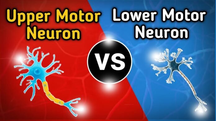
If you are looking for the difference between upper and lower motor neurons, you have come to the right place!
This article will discuss the distinction between upper and lower motor neurons. Motor neurons in the upper motor cortex transmit nerve impulses from the central nervous system to the lower motor cortex, which transmits them to the muscles. Both upper and lower motor neurons comprise the somatic nervous system, which controls voluntary muscle movements. The biological functions of upper and lower motor neurons are significantly different from one another.
Introduction
The lower motor neuron is the motor component that connects with the muscles. In contrast, the upper motor neuron is part of the central nervous system that controls movement and transmits brain impulses to synapses on lower motor neurons.The motor part of the somatic nervous system is made up of upper and lower motor neurons. They are in charge of activating voluntary muscle movement. Voluntary muscular activities are started and coordinated in the motor cortex, which is located towards the back of the brain’s frontal lobe.
The Central Nervous System
Upper motor neurons are higher up in the Central Nervous System (CNS), while lower motor neurons are found in the lower areas of the CNS. The CNS is made up of the spinal cord and brain. A motor neuron, sometimes referred to as a motoneuron, is a type of neuron with a cell body in the motor cortex, spinal cord, or brainstem and an axon fibre that extends to the spinal cord or elsewhere (to directly or indirectly regulate the effector organs such as muscles and glands). Upper and lower motor neurons communicate with one another through the spinal cord.
Upper Motor Neuron
The cerebral cortex’s motor area or the brainstem is where the upper motor neuron, a specific type of motor neuron, begins. It is in charge of sending nerve impulses from the brain to the lower motor neurons. As a result, it is not engaged in transmitting nerve impulses to the muscles. A neurotransmitter known as glutamate sends nerve impulses from top motor neurons to lower motor neurons through glutamatergic receptors.
The six routes that make up the upper motor tract are the corticospinal tract, corticobulbar tract, colliculospinal tract, rubrospinal tract, vestibulospinal tract, and reticulospinal tract.
Lower Motor Neuron
The effector’s muscles receive nerve impulses from the higher motor neurons through the lower motor neuron. It could originate from the brainstem, the anterior grey matter, the anterior nerve roots, or the cranial nerve nuclei. The major role of lower motor neurons is to link the brainstem or spinal cord to the muscles. The lower motor neurons are hence the cranial and spinal nerves.
Types of Lower Motor Neurons
α-Motor Neurons
- The particular subset of LMNs that, when harmed, result in the recognisable clinical symptoms of an LMN syndrome is known as -motor neurons. These neurons’ cell bodies are somatotopically organised and originate in laminae VIII and IX of the ventral horn of the spinal cord.
- In other words, neurons that innervate axial muscles are found ventral to those that innervate extensor muscles. In contrast, neurons that innervate distal musculature are found lateral to those that innervate axial muscles.
- The contraction of the muscle fibres they innervate is the purpose of -motor neurons. Since -motor neurons are necessary for muscle contraction, it has been called “the final common pathway”.
- Since -motor neurons make up the efferent portion of the reflex arc, this can be done under conscious control by UMNs or by inducing the myotatic stretch reflex. Therefore, if the -motor neurons are inactive, there cannot be coordinated muscle contraction.
γ-Motor Neurons
- γ-motor neurons play a crucial role in maintaining nonconscious proprioception and controlling muscle tone. Even though γ-motor neurons are included in the LMN category, only α-motor neurons can be damaged and cause an LMN syndrome.
- In the ventral horn of the spinal cord, laminae VIII and IX also give rise to γ-motor neurons. These innervate the skeletal muscle fibres that make up the contractile spindles.
- Furthermore, while α-motor neurons receive input from muscle spindle Ia sensory afferents and UMNs, γ-motor neurons are entirely controlled by UMNs. These fibres are crucial for communicating a muscle’s length and speed.
- By making the polar ends of the fibre contract, γ-motor neurons maintain the fibre’s tautness. The tension in these fibres must be maintained for muscle spindles to maintain their sensitivity to muscular strain.
Upper vs Lower Motor Neuron Lesion
An upper motor neuron lesion develops in the neural pathway above the spinal cord’s anterior horn or the cranial nerves’ motor nuclei. In contrast, a lower motor neuron lesion affects the nerve fibres that run from the anterior horn of the spinal cord to the corresponding muscle.
Difference Between Upper and Lower Motor Neurons
|
Upper Motor Neurons |
Lower Motor Neurons |
| These neurons are found in the brain or brainstem. Alpha and Gamma motor neurons in the ventral horn are innervated. | These innervate the ventral horn of the spinal cord’s alpha and gamma motor neurons. |
| The spinal cord’s axons move downward. | Axons move radially to innervate muscles. |
| The Central Nervous System is the sole recipient of this motor system. The upper motor neuron regulates postures to provide a solid background upon which it is necessary to commence voluntary activity. It is also responsible for successfully controlling voluntary movement, maintaining muscular tone, sustaining the body against gravity, and regulating postures. | The peripheral nervous system’s (PNS) efferent neuron connects the CNS to the muscle that will get the innervation. It is a nerve cell that terminates in skeletal muscle and is found in the spinal cord. |
| UMNs perform a wide range of regulatory tasks. They can either directly (mono synaptically) or indirectly influence the activity of α- and γ-LMNs (via interneurons). | This neuron represents the CNS’s whole range of functions. |
| Upper motor neuron cell bodies are larger than lower motor neuron cell bodies. | Lower motor neurons have considerably smaller cell bodies. |
| These send motor commands from the brain to the lower motor neuron synapses. | These neurons gather the impulses sent from the upper motor neuron to the body’s muscles. |
| Multiple sclerosis, stroke, and spinal cord damages are among the illnesses associated with its dysfunction. Primary lateral sclerosis is a motor neuron illness exclusively affecting the upper motor neuron. | Body weakness, muscle fasciculation (twitching), and muscle atrophy are all caused by its loss. |
| These are categorised according to the routes they take. | The sort of muscle fibres they innervate determines how they are categorised. |
| These connect to the lower motor neurons through synapses. | These create synapses with the body’s muscles. |
| Various conditions, diseases linked with higher motor neurons cause the motor neurons in the cortex and tronco encefalico motor nucleus to degenerate. Weakness, spasticity, motor clumsiness, and hyperreflexia are symptoms. | The General Somatic Efferent includes all neurons that innervate striated voluntary skeletal muscle. Those fibres are here (derived from somites and somatic mesoderm in the limb buds of the wall and somitomeres in the head). All spinal and cranial nerves, except I, II, and VIII, contain lower m. |
Frequently Asked Questions (FAQs)
Q1. What distinguishes UMN and LMN from one another?
Ans: A lesion of the neural pathway above the motor nuclei of the cranial nerves or the anterior horn of the spinal cord is referred to as an upper motor neuron lesion. Those nerve fibres that pass from the anterior horn of the spinal cord to the accompanying muscle are affected by a lower motor neuron lesion (s).
Q2. What symptoms indicate LMN lesion?
Ans: Weakness, muscle atrophy (wasting), and fasciculations are indications of LMN injury (muscle twitching). Any muscle group, including the arms, legs, chest, and bulbar region, may exhibit these symptoms. In the case of classical ALS, a person will have UMN and LMN symptoms in the same area, such as an arm.
Q3. What two types of motor neurons are there?
Ans: Specialised brain cells called motor neurons are within the spinal cord and brain. Their two main groupings are the top and lower motor neurons.
Q4. What is LMN’s weakness?
Ans: Clinical symptoms of lower motor neuron (LMN) disorders include muscle atrophy, weakness, and hyporeflexia without sensory involvement. They may result from diseases that damage the motor axon, the anterior horn cell, or the myelin surrounding these structures.
Q5. What do upper motor neurons do?
Ans: First-order neurons called upper motor neurons are in charge of carrying the electrical impulses that start and regulate movement. Coordinating movement is done via several descending UMN tracts. The pyramidal tract is the main UMN tract that stimulates voluntary movement.
